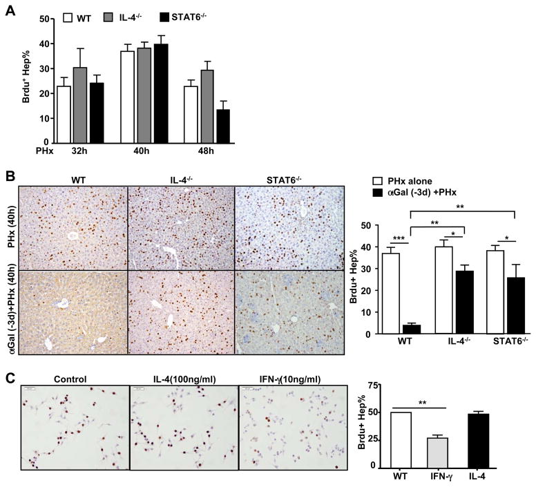
Fig. 4. The inhibitory effect of α-GalCer on PHx-induced liver regeneration is partially diminished in IL-4−/− and STAT6−/− mice.
(A) WT, IL-4−/−, and STAT6−/− mice were subjected to PHx, and liver regeneration was determined by BrdU incorporation. (B) Representative images of BrdU immunostaining from PHx mice treated with or without α-GalCer. The total number of BrdU+ hepatocytes is presented in the right panel. (C) Representative images of BrdU immunostaining from cultured AML12 cells treated with or without cytokines for 24h. The percentage of BrdU+ cells is presented in the right panel. Valuesin panels A–C represent the means ± SEM (n=8–10). *P<0.05, ** P<0.01; ***P<0.001

