
Figure 1.
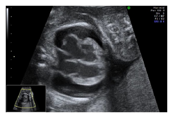
Fetal echocardiography at 21 weeks of gestation. A 2D image of the four-chamber view showed cardiomegaly, dilated ventricles, and atria with thickened and echo-dense walls, as well as massive pericardial effusion.
Official websites use .gov
A
.gov website belongs to an official
government organization in the United States.
Secure .gov websites use HTTPS
A lock (
) or https:// means you've safely
connected to the .gov website. Share sensitive
information only on official, secure websites.
Fetal echocardiography at 21 weeks of gestation. A 2D image of the four-chamber view showed cardiomegaly, dilated ventricles, and atria with thickened and echo-dense walls, as well as massive pericardial effusion.