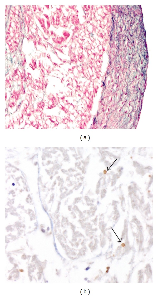
Figure 2.

Histopathology obtained during the infant autopsy. (a) Cardiac hematoxylin and eosin staining showed dilatation and massive endocardial fibroelastosis appearing as a thick white scale covering the inner surface of the left ventricle cavity. (b) The immunohistochemical analysis showed extensive inflammatory cell (arrow) infiltrates in the myocardium.
