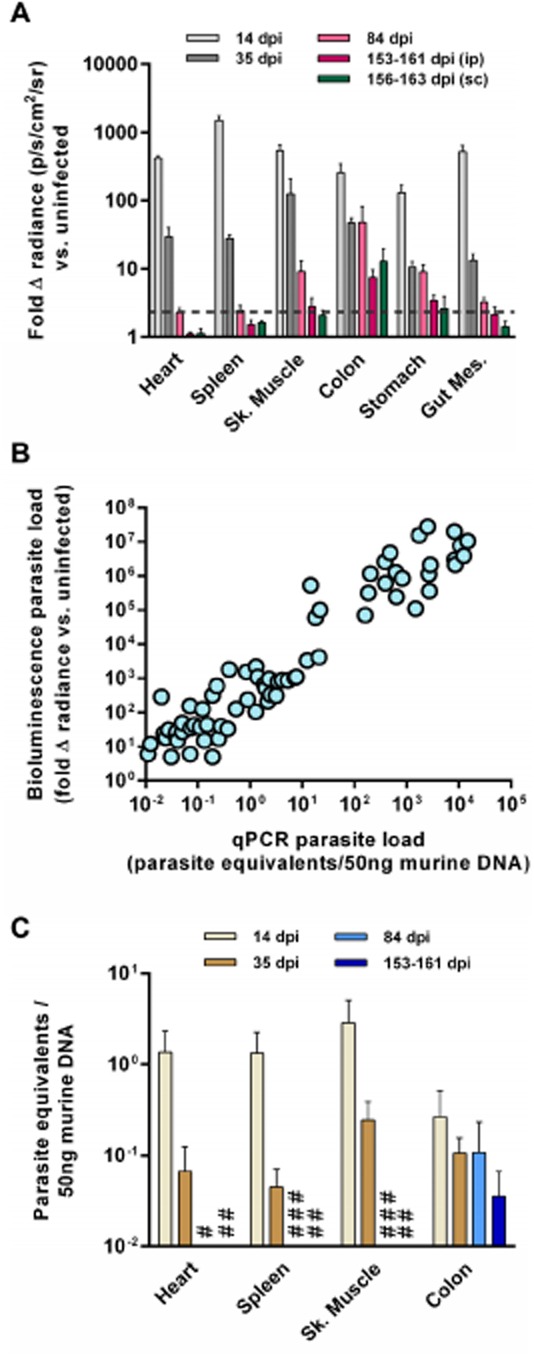
Quantification of tissue-specific parasite loads in
T. cruzi-infected BALB/c mice.A and B. Quantification of
ex vivo bioluminescence for selected organs and tissues taken immediately
post-mortem from BALB/c mice infected with
PpyRE9h luciferase-expressing
T. cruzi.
Animals were inoculated by i.p. injection of 1 × 10³ trypomastigotes and ex vivo imaging was performed at 14 dpi (n = 8), 35 dpi (n = 9), 84 dpi (n = 9) and 153–161 dpi (n = 8). Ex vivo imaging was also performed at 156–163 dpi on mice that had been inoculated by s.c. injection of 1 × 10³ trypomastigotes (n = 6). Data are means + SEM of the fold-change in bioluminescence intensity for organs from infected mice compared with matching organs from uninfected mice and are pooled from two independent experiments (n ≥ 3 mice per time point per experiment). Dashed line indicates the detection threshold equal to the mean +2SDs of the fold-change in bioluminescence intensity for empty regions of interest (ROI) in the images obtained for 153–160 dpi (ip) infected mice compared with empty ROI in the images obtained for uninfected mice.
Correlation between ex vivo bioluminescence-inferred and qPCR-inferred parasite loads in individual tissues samples (n = 70 samples from 20 mice from four independent experiments).
qPCR analysis of tissue-specific parasite burdens in mice inoculated i.p and sacrificed 14, 35, 84 and 153–161 dpi (n = 3–4 per group, one experiment). DNA was extracted from tissue samples and the relative amounts of T. cruzi DNA and murine DNA were quantified by real-time PCR amplification of a parasite locus (195 bp satellite) and a mouse control gene (gapdh). #, 84 dpi below detection limit; ##, 153–161 dpi below detection limit; ###, 84 dpi not done.

