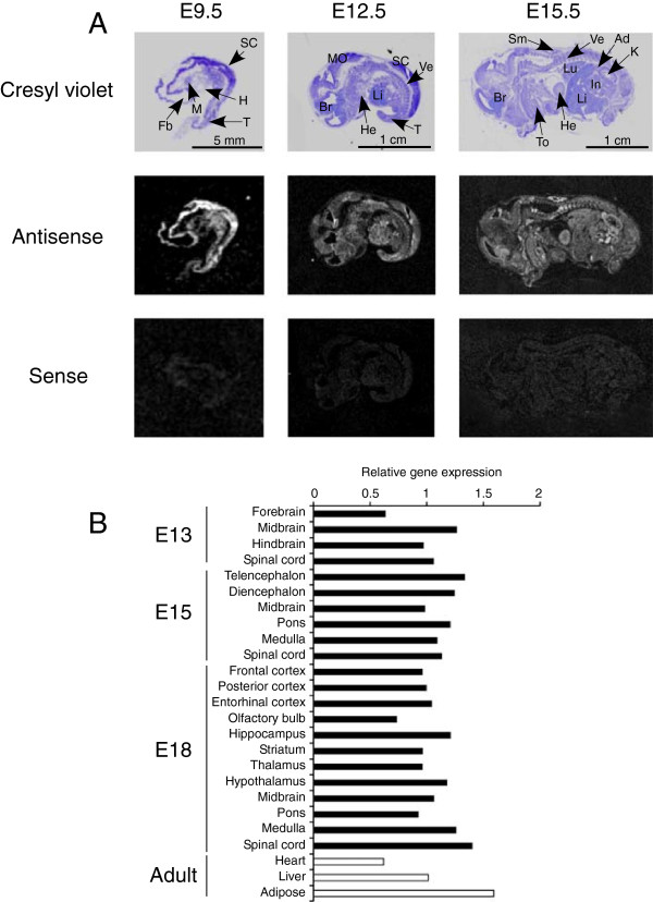
Figure 1.

LMF1 expression in the mouse embryo. (A)In situ hybridization of embryo sections with probes representing anti-sense (middle panels) and sense (bottom panels) transcripts of LMF1 at different days post coitus. The top panels show bright-field images of cresyl violet-stained sections. Ad, adrenal; Br, brain; Fb, forebrain; H, heart primordium; He, heart; In, intestine; K, kidney; Li, liver; Lu, lung; M, mandibular component of branchial arch; MO, medulla oblongata; SC, spinal cord; Sm, skeletal muscle; T, tail; To, tooth primordium; Ve, vertebrae. (B) Real-time PCR analysis of relative LMF1 expression in embryonic and adult tissues.
