
Figure 2.
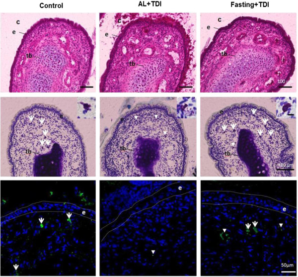
Representative photomicrographs of the distal end of the maxilloturbinate in TDI-sensitized nasal allergy model rat. Hematoxylin and eosin stainig (Upper). The mucus secretion in the nasal cavity and epithelia was increased in the AL with TDI. Toluidine blue staining (Middle). The degranulated mast cells presented in lamina propria of nasal epithelia in the AL with TDI. Insets show the magnified images of mast cell. Immunohistochemical detection of mast cell tryptase (Lower). Tb: turbinate bone. C: nasal cavity E: nasal epithelium. Arrow: non-degranulated mast cell. Arrow head: degranulated mast cell.