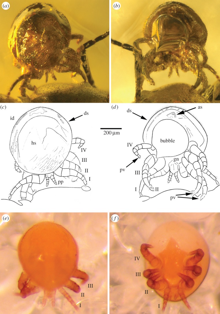
Figure 2.
Details of the fossil mite in (a) dorsal view and (b) ventral view, together with respective interpretative drawings (c,d) and comparative dorso-ventral images (e,f) of the living species Myrmozercon brevipes Berlese, 1902. as, anal shield; ds, dorsal setae; gn, gnathosoma; hs, holodorsal shield; id, idiosoma; pp, pedipalp; pv, pulvillus; legs numbered I–IV. (Online version in colour.)

