
Figure 2. 5-HT neurons express ERα.
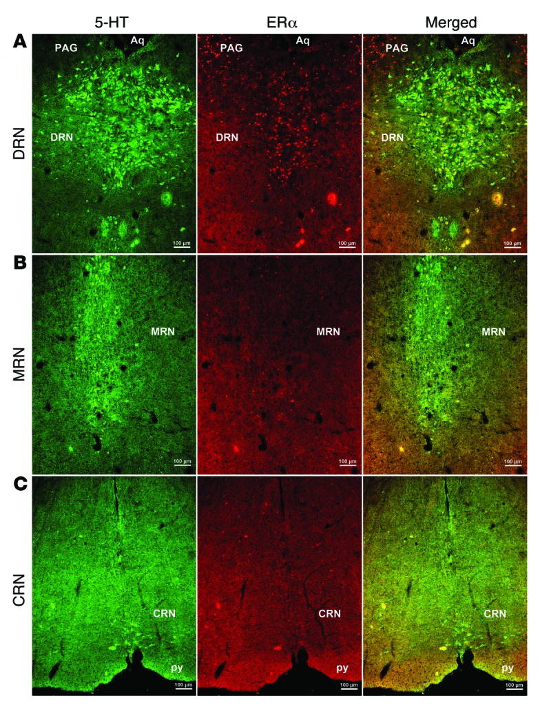
Representative immunofluorescent images for 5-HT (left, green) and ERα (middle, red) in coronal mouse brain sections containing the DRN (A), MRN (B), and CRN (C). Yellow neurons in the right panels indicate 5-HT neurons that coexpress ERα. Scale bars: 100 μm. Aq, aqueduct; PAG, periaqueductal gray; py, pyramidal tract.