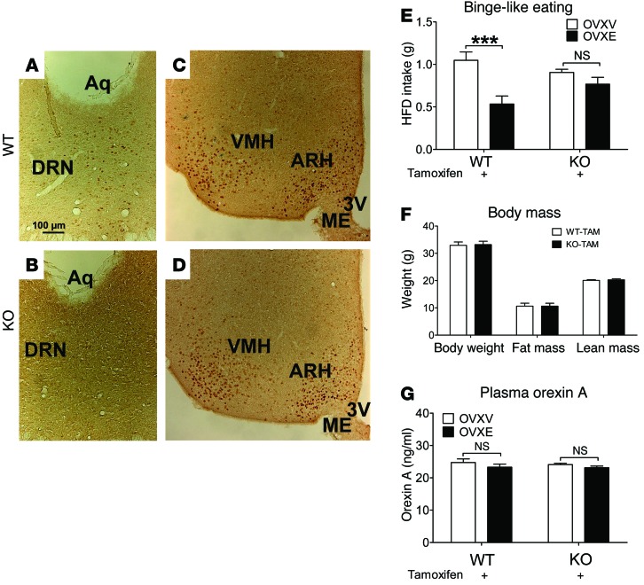
Figure 3. ERα in 5-HT neurons mediates estrogenic actions to inhibit binge-like eating in female mice.
(A–D) Representative immunohistochemistry for ERα from female WT (A and C) and KO (B and D) mice (after tamoxifen inductions). Scale bars: 100 μm. 3V, third ventricle; ME, median eminence. (E) WT and KO mice received tamoxifen inductions at 8 weeks of age (3 mg/injections, i.p., 24 hours apart). At 24 weeks of age, mice were ovariectomized and implanted with s.c. 17β-estradiol pellets (0.5 μg/d for 60 days; OVXE) or vehicle pellets (OVXV). After a 7-day recovery, mice were subjected to intermittent HFD exposure for 1 week, as described in Methods. At the end of that week, HFD and chow diet were provided to cages at 11:00 am, and 2.5-hour HFD intake was measured. n = 7–10/group. Results are shown as mean ± SEM. ***P < 0.001, between OVXV and OVXE mice in 2-way ANOVA analyses followed by post hoc Bonferroni’s tests. (F) Body weight, fat mass, and lean mass of WT and KO mice measured when binge-like behavior was assessed. n = 16 or 18/group. Results are shown as mean ± SEM. (G) Plasma orexin A measured in WT and KO mice after assessment of binge-like eating behavior. n = 6–7/group. Results are shown as mean ± SEM.

