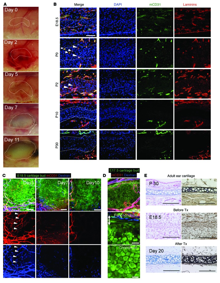
Figure 1. Rudimentary ear cartilage (mesenchymal condensation) is transiently vascularized in vivo.
(A) Gross observations of E18.5 CAG-EGFP murine immature cartilage transplants at multiple time points. (B) Immunohistological analyses of mCD31 and laminins in ear cartilage from P0 to P30. Dotted line indicates the border between the chondral and perichondral layers of the mouse ear cartilage. Scale bar: 50 μm. (C) Intravital confocal images of the same field of view at 3, 7, and 10 days after transplantation. Arrowhead indicates the vascularized area of the transplants. Endothelial cells and blood perfusion were visualized intravitally using Alexa Fluor 647–conjugated mCD31 antibody and fluorophore-conjugated dextran injected via the tail vein. Scale bars: 75 μm. (D) Formation of mature cartilage 20 days after transplantation. The developed chondral layer was composed of homogenous polygonal mature chondrocytes, whereas the perichondrium (double-headed arrow, outer fibrous layer) was composed of spindle-shaped putative progenitor cells that were proximate to blood vessels, similar to normal ear cartilage. Scale bars: 200 μm (top and middle panels) and 25 μm (bottom panel). (E) Terminal differentiation from murine progenitor cells into mature chondrocytes after transplantation into a cranial window. Alcian blue and EVG staining confirmed the formation of mature elastic cartilage from E18.5 immature cartilage transplants after 20 days compared with E18.5 and P30 cartilage sections. Tx; transplantation. Scale bars: 100 μm.

