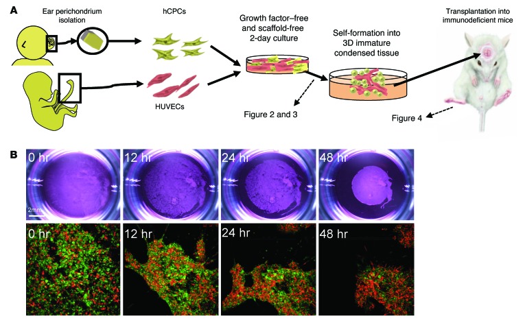
Figure 2. Self-driven condensation of hCPCs by endothelial cell coculture on a soft substrate.
(A) Schematic diagram of our culturing method. (B) Time-dependent changes in macroscopic and fluorescence microscopic images in vitro. hCPCs self-assembled into condensed 3D immature cartilage in vitro, without the aid of scaffolds or soluble factors. Green indicates HUVECs; red indicates human cartilage from CPCs. Bottom row original magnification: ×10.

