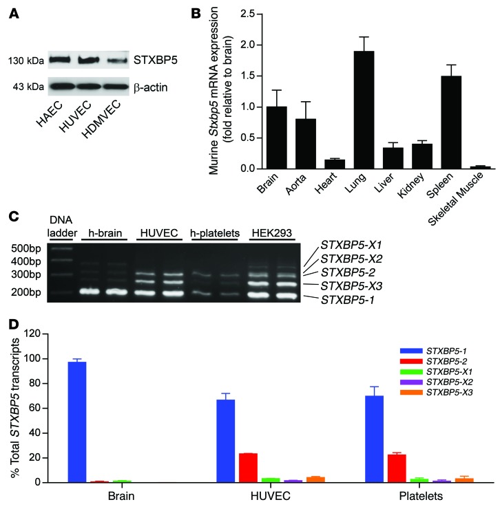
Figure 1. STXBP5 expression and transcript variants.
(A) STXBP5 expression in human ECs. Lysates of cultured HAECs, HUVECs, and HDMVECs were probed by IB with antibody against STXBP5. Blotting to β-actin was used as a loading control. Representative of 3 separate experiments. (B) Stxbp5 expression in murine tissue. RNA was isolated from WT mouse tissues, and Stxbp5 mRNA expression was measured by qPCR and normalized to mRNA level in brain (n = 3). (C) Human STXBP5 transcript variants. STXBP5 transcript variants were detected by RT-PCR using primers flanking splice region in human brain, HUVECs, human platelets, and HEK293 cells. The products were separated by agarose gel, sequenced, and compared with NCBI reference sequences (STXBP5-X1, PCR product 370 bp in length, 99% similarity to NCBI entry; STXBP5-X2, 322 bp, 95% similarity; STXBP5-2, 307 bp, 100% similarity; STXBP5-X3, 262 bp, 100% similarity; STXBP5-1, 199 bp, 100% similarity). (D) Relative abundance of STXBP5 transcript variants in human brain, HUVECs, and human platelets, measured by qPCR using variant-specific Taqman probes and expressed relative to brain STXBP5-1 (assigned as 100%) (n = 3). All data are mean ± SD.

