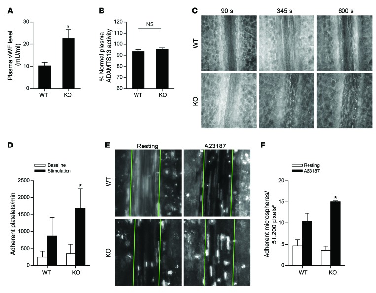
Figure 5. STXBP5 inhibits endothelial exocytosis in mice.
(A) Higher plasma levels of vWF in Stxbp5 KO than WT mice. Plasma vWF levels were measured by an ELISA from retroorbital bleeding (n = 8~12). (B) Similar plasma ADAMTS13 activity in Stxbp5 KO and WT mice. ADAMTS13 activity in mouse plasma was measured by a fluorescent assay and expressed as percentage of that in pooled WT plasma. (C) Intravital microscopy of platelet-EC interactions in vivo. Platelets were fluorescently labeled in WT and Stxbp5 KO mice. 10 μM A23187 was superfused onto the mesenteric venule after 120-second baseline recording. Platelet adhesion to the mesenteric venule was continuously recorded by intravital microscopy. Shown are representative images at baseline (90 seconds) and after A23187 stimulation (345 and 600 seconds). Original magnification, ×20. (D) Stxbp5 deficiency increases platelet-EC interactions in vivo. Quantification of platelet adhesion in C, calculated by averaging the number of platelets that remained transiently within at least 2 consecutive frames, and expressed as adherent platelets per minute in a 51,200-pixel2 area (n = 6 mice per genotype). (E) Stxbp5 deficiency increases microsphere-EC interactions in vivo. Mice were perfused with fluorescent microspheres conjugated with antibody against P-selectin, and adhesion of microspheres was imaged 2 minutes before (resting) and 8 minutes after A23187 superfusion on mesenteric venule. Green lines denote the borders of the vessel within which adherent microspheres were counted. Original magnification, ×20. (F) Quantification of adherent microspheres in E (n = 3–4 mice per genotype). All data are mean ± SD. *P < 0.05 vs. WT.

