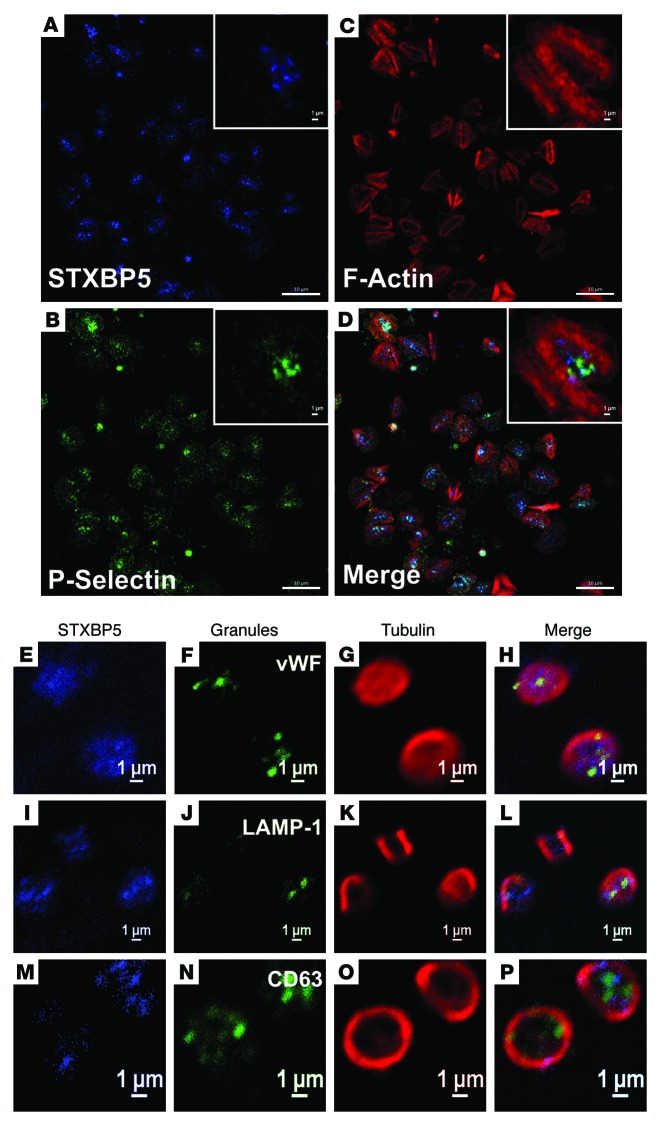
Figure 3. STXBP5 is present in platelets.
Washed human platelets were allowed to bind to fibrinogen-coated coverslip for 1 h at 37°C. After washing away the unbound platelets, the bound platelets were fixed and immunostained for STXBP5 (A), P-selectin (B) and F-actin using TRITC-phalloidin (C). Platelets were additionally stained with anti-STXBP5 antibodies (E, I, and M), anti-tubulin antibodies (G, K, and O) and antibodies against markers for α granules (vWF; F), lysosomes (LAMP-1; J), and dense granules (CD63; N). Images in A–D were taken using a Zeiss LSM 780 confocal microscope and processed using ZEN blue software, and images in E–P were taken using a Nikon A1R confocal microscope and processed using NIS-Elements AR 3.2 software; D, H, L, and P show 3-color merged views. Scale bars: 10 μm (A–D); 1 μm (A–D, insets, and E–P).

