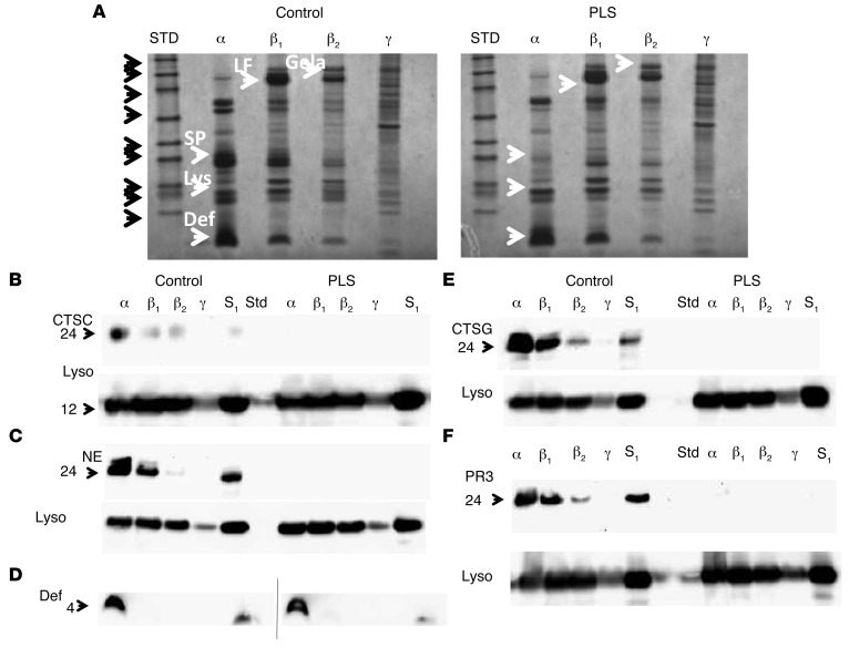
Figure 1. Deficiency of cathepsin C and serine proteases in PLS neutrophils.
(A) Coommassie-stained 4%–12% SDS-PAGE–separated proteins from isolated azurophil granules (α), specific granules (β1), gelatinase granules (β2), and light membranes (γ) from PLS neutrophils and neutrophils from a normal control. Molecular weight (MW) standards are indicated with black arrows: from top, 225; 102; 76; 52; 38; 31; 24; 17; 12 kDa. Selected proteins are indicated by white arrows. SP, serine proteases; Lys, lysozyme; Def, defensins; LF, lactoferrin; Gela, gelatinase. (B–F) Western blotting for proteases of the isolated fractions α, β1, β2, and γ and of the postnuclear supernatant (S1), which is the material loaded onto the density gradient. (B) CTSC and lysozyme (Lyso); (C) NE and lysozyme; (D) defensin (control and patient samples were run on 2 separate gels that had a slightly curved run); (E) CTSG and lysozyme; (F) PR3 and lysozyme. Blotting was done first for serine proteases. The blots were then stripped and reprobed with antibody against lysozyme as a loading control.

