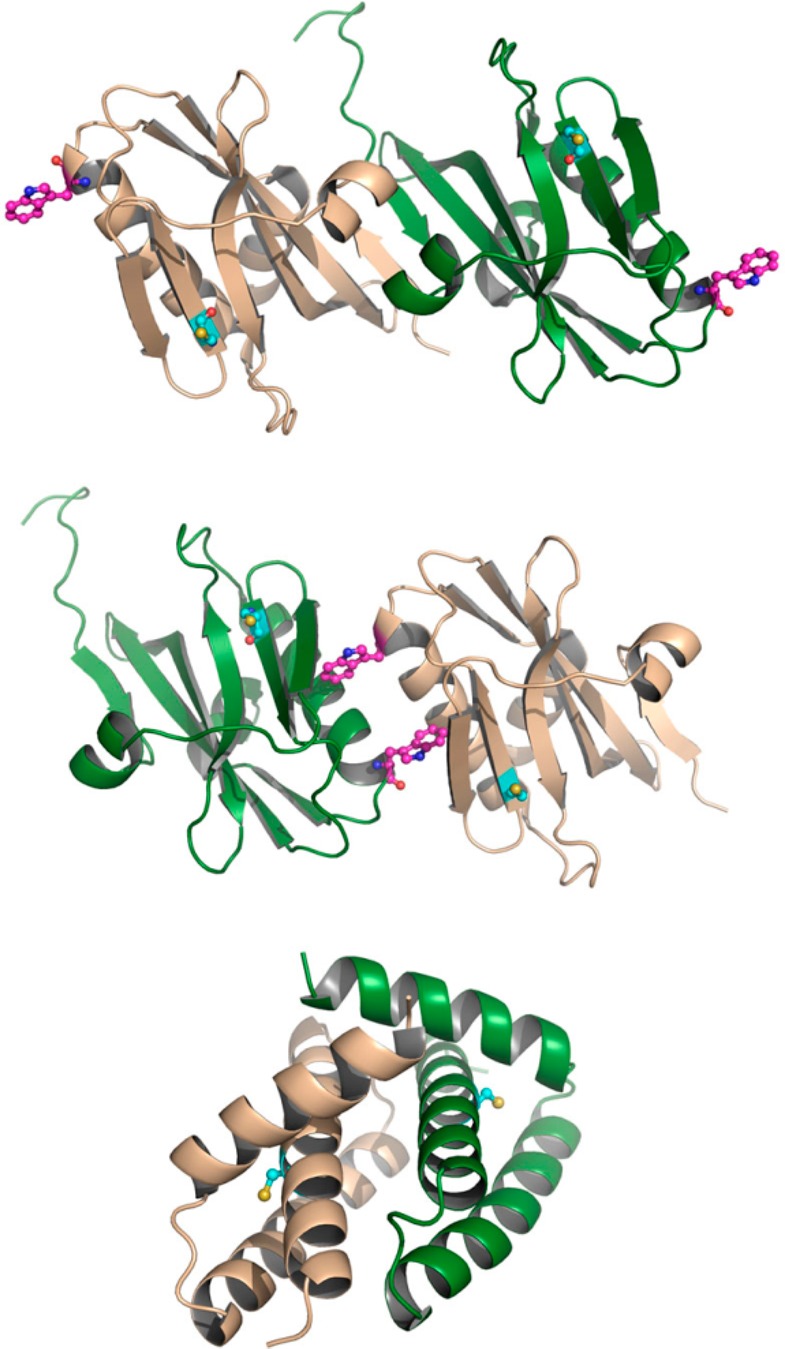
Figure 1.

Dimerization states of the domains of NS1: (top) strand–strand ED dimer; (middle) helix–helix ED dimer; (bottom) RBD dimer. Protein shown as green and beige cartoon. In the top and middle parts, residues Cys116 and Trp187 are shown as ball-and-stick in cyan and magenta, respectively. In the bottom part, Cys13 is shown as ball-and-stick in cyan.
