
Figure 1.
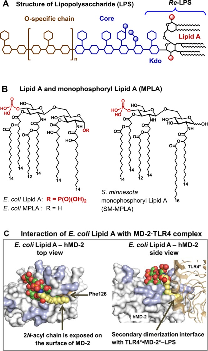
(A) Structure of LPS, with Re-LPS and Lipid A. (B) Structures of TLR4 agonist E. coli Lipid A and MPLA. (C) Co-crystal structure of E. coliRa-LPS-hMD-2·TLR4 (PDB code: 3FXI; only Lipid A portion of LPS is shown for clarity), top and side views. Phe126 (orange) together with 2-N-acyl chain (yellow) creates a hydrophobic patch at the dimerization interface with the second TLR4*·MD-2* complex (brown). Positively charged Arg and Lys (blue) at the rim of the binding pocket of MD-2 are involved in the ionic interactions with the Lipid A phosphates. Images were generated with PyMol.