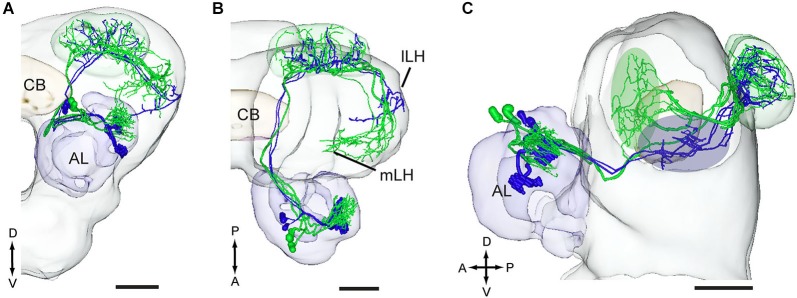
Figure 9.

Three-D models of pheromone neurons (green) and plant odor neurons (blue) registered into the standard brain model of the H. virescens male. (A) Frontal orientation. (B) Dorsal orientation. (C) Sagittal orientation. The green and blue ovals indicate the different target regions of the two neuron categories in the lateral horn. lLH, lateral part of the lateral horn; mLH, medial part of the lateral horn. A, anterior; D, dorsal; L, lateral; P, posterior. Scale bars, 100 μm.
