Figure 2.
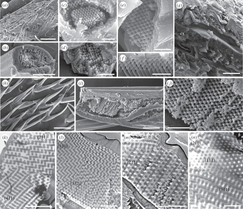
Electron micrographs of fossil (a–g,k,l) and modern (h–j,m,n) scales. (k–n) are transmission electron micrographs; all other images are scanning electron micrographs. (a,h) Surface of the fossil cuticle showing alignment of scales. (b–f,i,j) Detail of fractured scales showing 3DPCs in the scale lumen; (f) is an ion milled vertical section. (g) Fractured section through the cuticle of the fossil weevil showing fine lamination in the exocuticle (ex) and bundles of chitin microfibrils in the endocuticle (en); sc, scale. (k–n) Transmission electron micrographs of vertical sections of fossil (k,l) and modern (m,n) scales showing photonic polycrystallite domains. Numbers in parentheses denote distinctive motifs characteristic of specific planes of the single diamond morphology. Scale bars, (a) 50 µm; (b) 10 µm; (c–f,k–n) 1 µm; (g,i) 5 µm; (h) 25 µm; (j) 2 µm.

