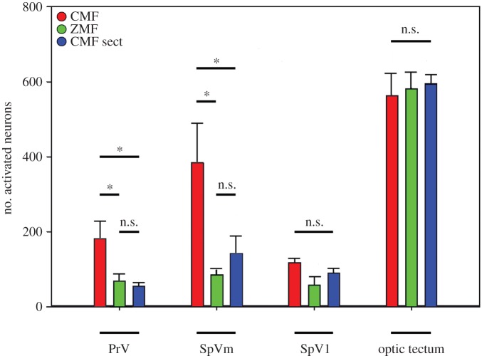
Figure 3.
Quantification of ZENK-activated neurons in PrV, SpV, and the optic tectum of all investigated pigeons. Birds experiencing a CMF are shown in red, birds experiencing a zero magnetic field (ZMF) are shown in green, and birds with sectioned V1 experiencing CMF conditions (CMF sect.) are shown in blue. Since we counted ZENK-positive neurons in every second slide bilaterally throughout the relevant areas, the numbers indicated on the y-axis reflect the approximate absolute number of ZENK-activated neurons in one side of brain, or when multiplied by 2, the total number of bilaterally activated neurons in PrV and SpV. Error bars indicate s.e.m.

