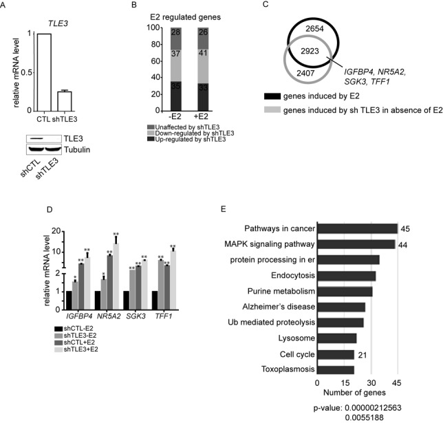
Figure 1.

TLE3 is involved in the regulation of ERα target genes transcription. (A) RT-qPCR and western blot analysis of MCF-7 cells infected with lentiviruses expressing no (shCTL) or one shRNA (shTLE3) directed against TLE3. TLE3 mRNA expression was normalized to GapDH and Tubulin was used as the leading control for the western blot. (B) Histogram representing the distribution of genes affected by the knockdown of TLE3. The genes regulated by E2 were compared to the genes up- and down-regulated by shTLE3 in absence or in presence of E2. (C) Venn diagram of genes up-regulated by E2 and genes up-regulated by the knockdown of TLE3 in absence of E2. (D) MCF-7 cells were infected with an empty lentivirus (shCTL) or a lentivirus carrying an shRNA against human TLE3 (shTLE3) and treated with vehicle (-E2, ethanol 0.1%) or 17β-estradiol (+E2 10−7M) for 3 h. Total RNA was used to generate cDNA for RT-qPCR. The results presented are average of at least three independent experiments (Student's t-test * = P < 0.05; ** = P < 0.01) (E) Gene ontology of the overlapping genes reported in the Venn diagram in (C). er: endoplasmic reticulum; Ub: ubiquitin.
