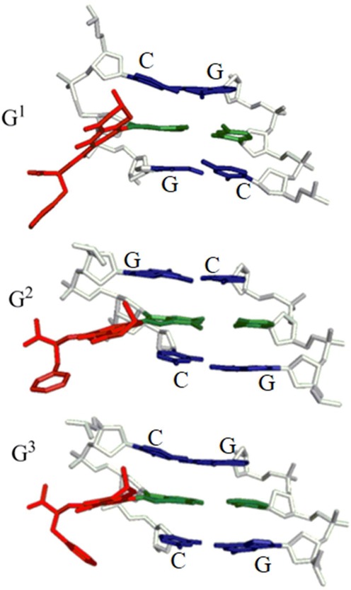
Figure 3.

Representative structures of the major groove conformation of adducted DNA with the neutral OTB-dG adduct (α-rotamer) incorporated at the G1, G2 or G3 position in the NarI recognition sequence. Central trimers are shown that include the lesion–base pair (green, with bulky moiety in red) and the flanking base pairs (blue) viewed from the major groove side. The sugar-phosphate backbone is in white and hydrogen atoms are removed for clarity.
