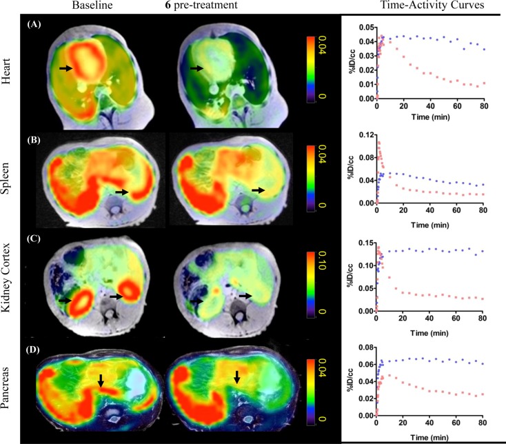
Figure 3.
[11C]6 PET-MR imaging. Axial views of summed PET images (40–80 min) superimposed with MR images from the same baboon following injection of radiotracer (4 mCi/baboon). Images illustrate tracer uptake in organs of interest at baseline and after pretreatment with unlabeled 6 (0.5 mg/kg). Robust blocking was observed in organs of interest including: A, heart; B, spleen; C, kidneys; and D, pancreas. Time–activity curves (baseline, blue; blocking, red) demonstrate a high specific binding of [11C]6 in these peripheral organs as the percent injected tracer dose per cm3 tissue is markedly reduced by blocking.

