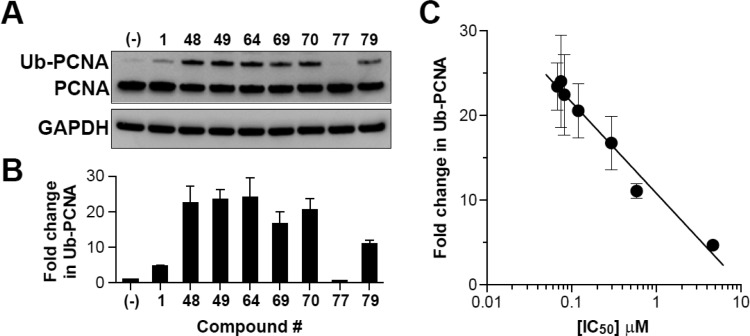
Figure 2.
Disruption of USP1/UAF1-catalyzed deubiquitination of PCNA in H1299 cells. (A) H1299 cells were treated with the indicated compounds at 20 μM for 4 h. Whole cell extracts were separated on 4–12% gradient SDS-PAGE and subjected to Western blotting with antibodies against PCNA and GAPDH (loading control). (B) Fold changes in monoubiquitinated PCNA (Ub-PCNA) were calculated by normalizing to unmodified PCNA and GAPDH. The data represent the mean ± SEM of two biological replicates. (C) Correlation between mean IC50 values obtained in the HTS assay and fold change in Ub-PCNA in H1299 cells (Pearson r = −0.988; P < 0.0001).

