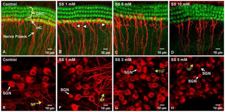
Fig. 5.

Cochlear organotypic cultures from postnatal day 3 rats labeled with Alexa-488 conjugated phalloidin to identify F-actin that is heavily expressed in the stereocilia and cuticular plate of outer hair cells (OHC) and inner hair cells (IHC). Nerve fibers (NF) and spiral ganglion neurons (SGN) labeled with a monoclonal antibody against class III –tubulin and Cy3-conjugated secondary antibody (red). (A-D) Control culture and cultures treated with escalating doses of SS for 48 h. Note loss of NF as SS dose increases; arrowhead points to NF. (E-H) Photomicrographs show SGN and NF from postnatal day 3 cochlear cultures. Note large round SGN and nerve fibers (NF) emanating from the soma of control cultures. Treatment with escalating doses of SS for 48 h results in loss of NF and shrinkage of SGN. (From Wei et al., 2010 with permission).
