
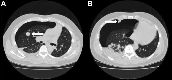
Figure 3.
CT scans of patient 3. Routine follow-up CT scan showing the pneumothorax on the right side. A shows one of the pulmonary metastasis, B shows a pleural metastasis.
Official websites use .gov
A
.gov website belongs to an official
government organization in the United States.
Secure .gov websites use HTTPS
A lock (
) or https:// means you've safely
connected to the .gov website. Share sensitive
information only on official, secure websites.
CT scans of patient 3. Routine follow-up CT scan showing the pneumothorax on the right side. A shows one of the pulmonary metastasis, B shows a pleural metastasis.