
Figure 5.
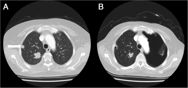
CT scans of patient 5. A: CT scan before the start of pazopanib showing multiple metastases in the right lung, one is indicated by the arrow. B: Routine follow-up CT scan during pazopanib treatment showing the left-sided pneumothorax and the earlier mentioned metastasis in the right lung is now showing cavitation.