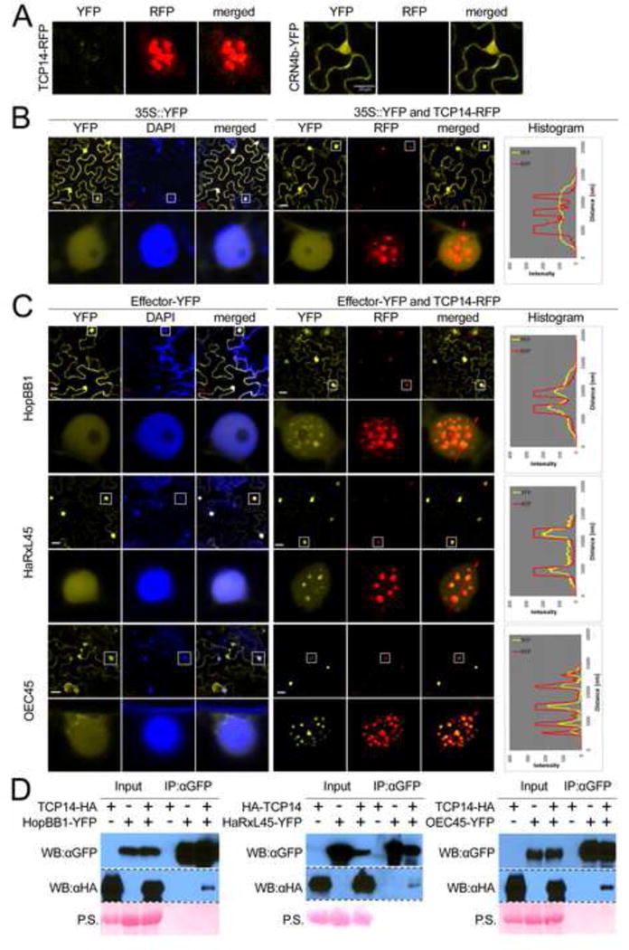
Figure 4. TCP14 re-localizes effectors to sub-nuclear foci.

A. Technical control demonstrating that YFP- and RFP channels do not leak into each other. The images show localization of TCP14-RFP and CRN4b-YFP; image data for YFP and RFP channels were collected for both. The same settings were then applied to all assays below. Note that TCP14-YFP forms sub-nuclear foci. B and C. The lower panel exhibits an enlarged view of a representative nucleus boxed in the upper panel. The histogram illustrates the intensity of fluorescent signal across the path indicated by the red arrow. All confocal pictures were taken 40–48 h after infiltration of Agrobacterium strains expressing the different fluorophore–tagged proteins. B. Negative control: TCP14 does not re-localize YFP. C. TCP14 re-localizes effectors from Psy (HopBB1), Hpa (HaRxL45) and Gor (OEC45) to sub-nuclear foci. D. TCP14 is co-immunoprecipitated by HopBB1, HaRxL45, and OEC45. All proteins were expressed from the CaMV 35S promoter in N. benthamiana leaves. ‘P.S’ in D denotes Ponceau S staining. See also Figure S4 and Table S4.
