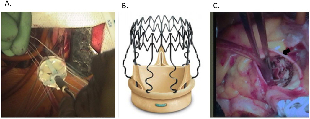
Figure 4.
Traditional sutured prosthetic aortic valve placement through a minimally invasive J-hemisternotomy (A). Percival S self-expanding aortic tissue valve with Sinus of Valsalva struts (B, from www.sorin.com/product/perceval-s). Perceval S valve placed in the aortic position (C, arrow).

