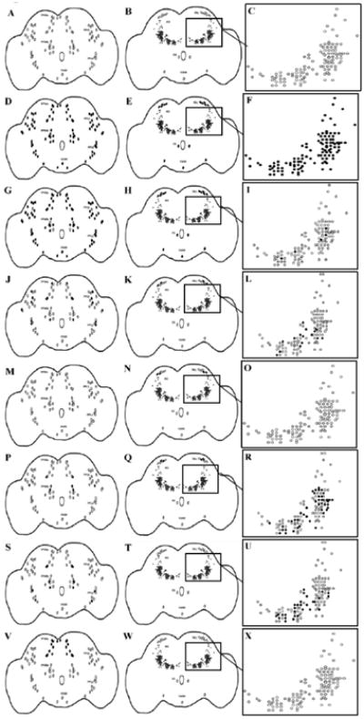
Figure 1.

Cartoon depictions of the Gal4 expressing neurons found to overlap with tyrosine hydroxylase-expressing neurons in a series of Gal4 lines. Column 1, focus posterior, column 2, focus anterior, column 3, focus AG magnified to show individual neurons. Black filled neurons represent those expressing both Gal4 and tyrosine hydroxylase, whereas open neurons represent those expressing only tyrosine hydroxylase. (A-C) a parental control line (elavC155/w1118); (D-F) a panneuronal GAL4 (elavC155); (G-I) THGal4; (J-L) 23y Gal4; (M-O) 103y Gal4; (P-R) 201y Gal4; (S-U) 4669 Gal4; (V-X) 854 Gal4. The pattern of overlap is bilaterally symmetrical. PPM (protocerebral posterior medial), PPL (protocerebral posterior lateral), EG (esophageal), VUM (ventral unpaired medial), PAL (protocerebral anterior lateral), AG (antennal glomeruli). n = 42 to establish the normal pattern of dopamine neurons in wild-type (CSwu) animals. n = 12-22 to determine overlap between tyrosine hydroxylase and Gal4 expression.
