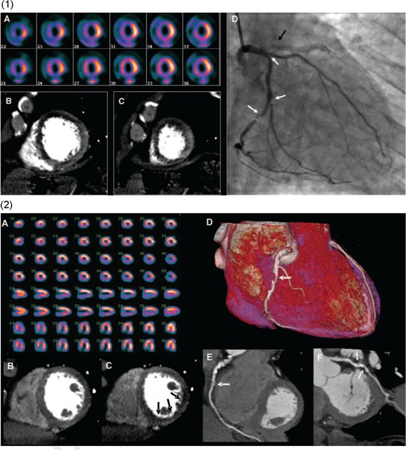
Figure 1.
(1) Images from a 64-row detector CTP. (A) A partially reversible perfusion deficit in the territory of the LAD and a primarily fixed perfusion deficit in the inferior wall on radionuclide myocardial perfusion imaging with increased subdiaphragmatic tracer uptake (stress, upper panels; rest, lower panels). (B and C) Adenosine stress CTP shows a dense perfusion deficit in the LAD territory and a subendocardial perfusion deficit in the inferior and lateral walls. (D) An invasive coronary angiogram demonstrates a left-dominant system with a totally occluded LAD (black arrow) as well as intermediate- and high-grade stenoses in a large ramus intermedius, the body of the left circumflex, and the ostium of the obtuse marginal artery (white arrows). (2) Images from a 256-row detector CTP. (A) A partially reversible perfusion deficit in the inferior and inferolateral wall on radionuclide MPI in this patient with exertional angina (stress, upper panels; rest, lower panels). Rest (B) and stress (C) CTP imaging shows a reversible subendocardial perfusion deficit in the inferior and inferolateral walls. Noninvasive angiography confirms a significant stenosis (white arrows) in the proximal right coronary artery (D and E) and the proximal left circumflex artery (F).27 (Color version of figure is available online.)

