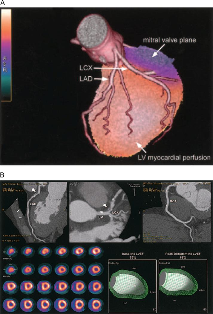
Figure 4.
(A) Fused 3D reconstructions of a coronary arteriogram and stress myocardial perfusion obtained in the same setting, assessed through integrated PET/CT. A full-motion cine can be viewed in the online-only Data Supplement Movie V. LCX indicates left circumflex artery; LAD, left anterior descending coronary artery.60 (B) An integrated PET/CT study. The CTA images demonstrate a noncalcified plaque (arrowhead) in the proximal left anterior descending coronary artery (LAD) with 50%-70% stenosis; however, the rest and peak dobutamine stress myocardial perfusion PET study (lower left panel) demonstrates only minimal inferoapical ischemia. In addition, LVEF was normal at rest and demonstrated a normal rise during peak dobutamine stress. Full-motion cines can be viewed in the online-only Data Supplement (Movies VI and VII). Ant, anterior; Endo Epi, endocardial plus epicardial; Inf, inferior; LCX, left circumflex; LM, left main; RCA, right coronary artery.60

