
Fig. 2.
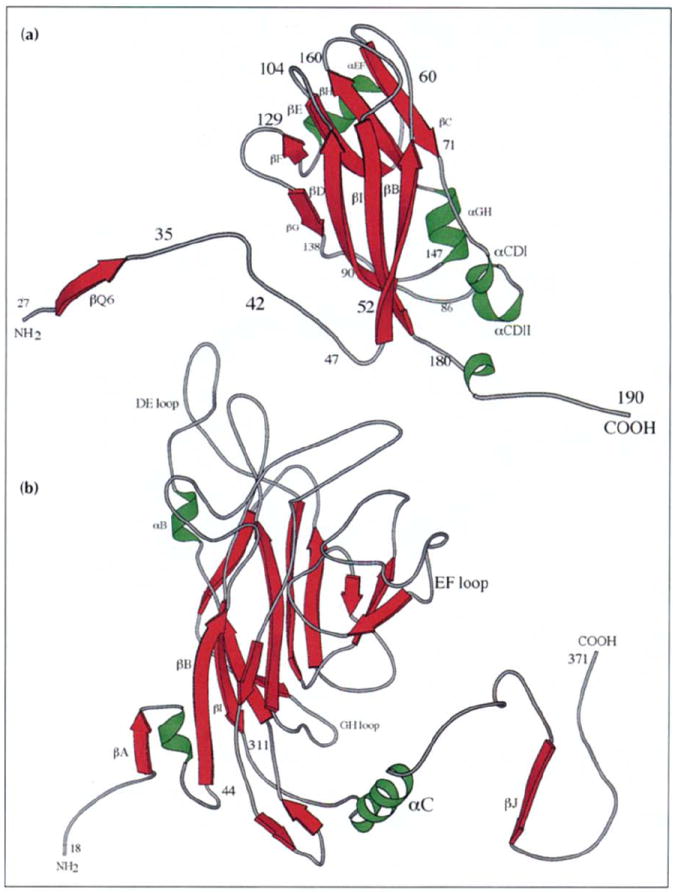
Ribbon diagrams showing the tertiary structure of the (a) CCMV and (b) polyoma virus VP1 capsid subunits (VP1 drawn at smaller scale). Virus exteriors are at the top of each diagram. Selected residues and secondary structure elements are labeled. (Polyoma virus coordinates were kindly provided by Thilo Stehle and Steven Harrison, Harvard University.)