
Fig. 7.
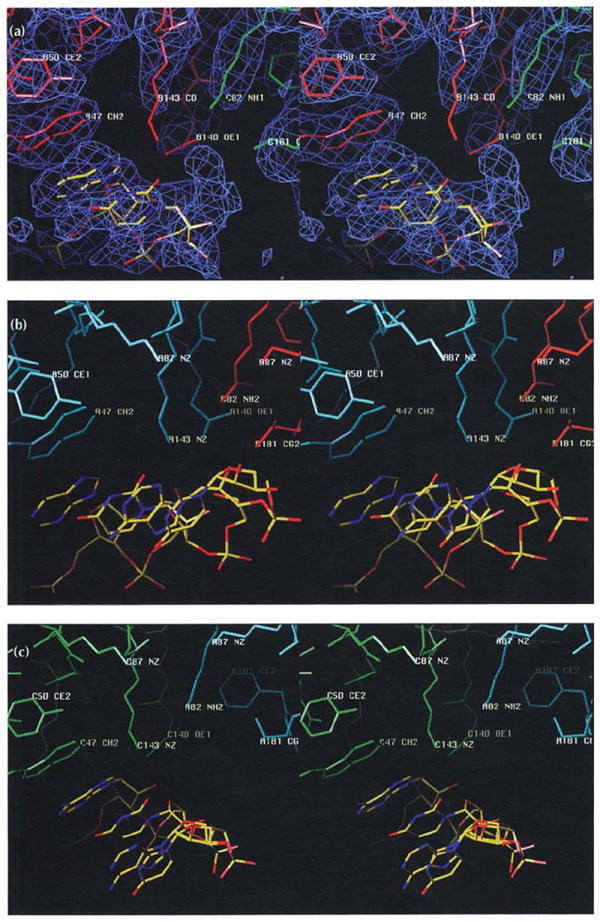
Similarly oriented stereoviews of the protein–RNA interactions at the A (blue), B (red), and C (green) subunit interfaces. The views are tangential to the protein shell looking away from the quasi three-fold axis along the subunit interfaces. The RNA model is colored according to atom type (nitrogen, blue; oxygen, red; and carbon, yellow). Selected residues are labeled as in Fig. 3, which shows the location of residues that interact with RNA. (a) The B-C subunit interface. Electron density (blue) at approximately 2σ contour level is shown for this interaction, but is left out of the following parts of the figure for clarity. (b) The A-B subunit interface. (c) The C-A subunit interface.