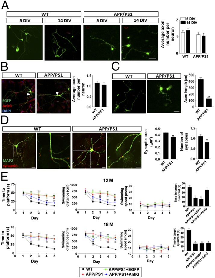
Fig. 4.
Impaired AIS filtering induced axonal, synaptic, and cognitive defects in APP/PS1 neurons and mice. (A) No difference was detected in the number of axons per neuron in 5 DIV and 14 DIV WT and APP/PS1 neurons. (Scale bar: 20 μm.) (B) No difference was detected in the number of axons per neuron in 21 DIV WT and APP/PS1 neurons. EGFP (green) was randomly transfected into neurons, and AnkG (red) was stained by its antibody. The arrow indicates the AIS. (Scale bar: 20 μm.) (C) The axons of the WT neurons were significantly longer than those of the APP/PS1 neurons at 14 DIV. (Scale bar: 20 μm.) (D) The number of synapses and overall synaptic area were remarkably larger in WT neurons than in APP/PS1 neurons at 14 DIV. Data are mean ± SE (n = 10 for each group). **P < 0.01 compared with WT. (Scale bar: 20 μm.) (E) The AnkG construct was packaged into adenovirus and infected in the hippocampal CA1 region of APP/PS1 mice. Overexpression of AnkG significantly improved Morris water maze spatial learning and memory performance in 12-mo-old, but not in 18-mo-old, APP/PS1 mice. Data are mean ± SE (n = 8 for each group). ##P < 0.01 compared with APP/PS1 or control; **P < 0.01 compared with WT.

