Fig. 2.
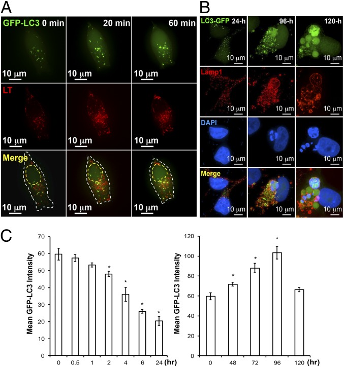
Prolonged ADI-PEG20 treatment can induce giant-autophagosome formation and affect autophagic flux pattern. (A) Representative time-lapse images showing ADI-PEG20 treatment induces autophagy and facilitates the fusion of autophagosome (green) and lysosome (red, LysoTracker) into autophagolysosome (yellow). The outline is shown by a dashed white line. The estimated nucleus location is shown by a dashed yellow line. (B) Prolonged ADI-PEG20 treatment induces abnormally sized autophagosomes (green), which colocalize with lysosome (red) and leaked DNA (blue). (C) ADI-PEG20 treatment induces autophagic flux with distinct kinetics. Mean GFP-LC3 intensity in each group was plotted as a bar graph. Data were collected from three independent experiments and are shown as mean ± SD; *P < 0.05.

