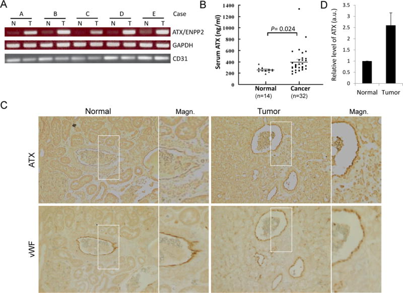
Figure 1.

Elevated ATX expression in endothelial cells (ECs) of tumor vessels, but not in tumor cells or in ECs of the normal kidney vasculature. A, expression of ATX (ENPP2) mRNA in endothelial cells of RCC and corresponding normal kidneys. ECs from human RCC specimens (T) and their normal counterparts (N) were isolated from a single-cell suspension using anti-CD31-coated magnetic beads. Total RNA was prepared and RT-PCR was performed for ENPP2, GAPDH, and CD31. B, serum ATX levels of RCC patients (Cancer) and healthy subjects (Normal) were measured by the enzyme-linked immunosorbent assay (ELISA). The difference in serum ATX levels between two groups was statistically significant (by Student’s t-test). C, immunohistochemical staining of RCC and normal kidney tissues. Human RCC specimens and their normal counterparts were stained using specific antibodies against autotaxin (ATX) and von Willebrand factor (vWF). A total of six samples were examined and a photograph of one representative case is shown. The right panels are magnified images of the areas boxed in the left panels (Magn.). D, densitometric analyses of endothelial ATX expression in normal and tumor kidney vasculature. The levels of immunohistochemically labeled ATX in vWF-positive vessels of RCC (Tumor) and their corresponding normal counterparts (Normal) from each of four patients were measured and expressed as means ± S.D. in arbitrary units (a.u.).
