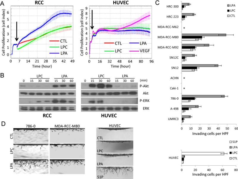
Figure 3.

Effects of LPA on cell proliferation, signaling and invasion of RCC and endothelial cells. A, HRC-223 (RCC) and HUVECs were seeded on E-Plates at 10,000 cells per well and continuously monitored for cell proliferation using The xCELLigence System. Arrowhead indicates the time point at which medium only (CTL), 20 μM LPC, 1 μM LPA, or 40 ng/ml VEGF was added. B, HRC-223 and HUVECs were serum-starved for 4 hours and treated with 20 μM LPC or 1 μM LPA for the indicated time points. Cell lysates were collected and analyzed by immunoblotting using the indicated antibodies. C-D, cell lines and primary cultures of RCC and HUVECs were induced to invade into 3D collagen matrices (2.5 mg/ml) under serum-free conditions in the absence (CTL) or the presence of 20 μM LPC, 1 μM LPA, or 1 μM S1P. After 48 hours, cultures were fixed, stained, quantitated for cell invasion (C), and photographed (D). Data are presented as mean numbers of invading cells per standardized field ± S.D. (n=4 fields) (*, no invasion). HUVECs were fixed and analyzed 24 hours after seeding.
