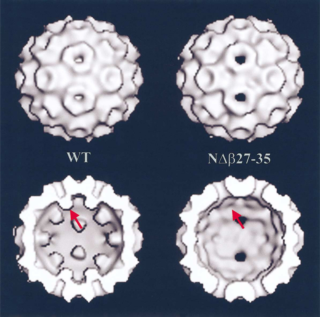
Fig. 3.
(Top) Shaded-surface representations of the image reconstructions of in vitro assembled CCMV wild-type (WT) and NΔβ27–35 empty particles. (Bottom) Same as (Top) but with front half of particles removed to show the particle interiors indicating the presence (WT) or absence (NΔβ27–35) of β-hexamer density at the pseudo-six-fold axes (arrow).

