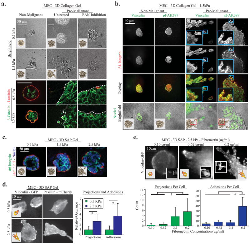
Figure One. In 3D, ECM cues promote vinculin recruitment to focal adhesions, projection formation, and cell invasion.

A. MEC spheroids composed of MCF10A cells in 0.5 kPa or 2 mg/ml 1.5 kPa 3D collagen gels for 24hrs. Invasion is increased by matrix stiffness and Ha-ras malignant transformation (MCF10AT cell line), and decreased via inhibition of integrin mediated FAK signaling (FAK Inhibitor 14 at 1μM). To document loss of normal 3D cell organization in stiff 3D collagen, cell polarity was studied 48 hours after seeding in collagen via immunostaining for β-catenin (cell-cell junctions) and pan-laminin (basal surface). Dotted white lines indicate spheroid edge. B. Pre-malignant and non-malignant MEC 1.5kPa 3D collagen gels for 48 hours, immunostained for integrin β1, vinculin and pFAK39. Images are maximum intensity z-projections of 1μm increment confocal image slices, and demonstrate focal adhesion formation at cell-ECM borders. C. Non-malignant MEC spheroids in 3D SAP gels with 0.8, 2.2 or 5mg/ml RADA-16 polymers; stained for polarity markers α6 Integrin (cell-cell junctions) and laminin-5 (basal polarity). D. Non-malignant MEC adhesions and projections in soft and stiff 3D SAP gels; with quantification of projections and adhesions per cell (p < .01; ±SD, n>18 cells per condition). E. Non-malignant MEC expressing vinculin-GFP In 3D SAP gels with controlled ECM fibronectin concentration. Images are maximum intensity z-projection of 1.0um confocal image planes. Cellular projections and adhesions per cell were significantly upregulated by ECM stiffness and fibronectin (p < .01; ±SD, n>12 cells per condition).
