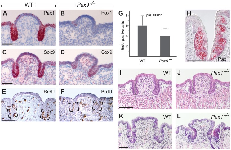
Figure 6. Pax1 and Sox9 are Pax9 targets in the proliferating compartment of the CVP trenches.
(A–F) Immunohistochemical staining on sections of the CVP at E15.5. (A,B) Pax1 is strongly expressed in the tips of epithelial trenches and in periderm cells covering the central dome of the wild type CVP (A), but not in the Pax9-deficient CVP (B). (C,D) Similarly, Sox9 expression is strongest in the epithelial trenches (C) and is barely detectable in the Pax9 mutant CVP (D). (E,F) BrdU-positive cells were counted in defined areas (boxed) of the CVP trenches from three wild type (n = 29 sections) and three Pax9 mutant CVPs (n = 28 sections). (G) The number of proliferating cells in the Pax9-deficient CVP is significantly reduced. Error bars illustrate standard deviation. (H) Pax1 immunostaining of one CVP trench in a 3 months old wild type mouse. (I,J) Morphology of the CVP at E18.5. The lengths of the CVP trenches (indicated by bars) were measured and shown to be reduced in the absence of Pax1 (for summary of measurements see Table S1). (K,L) Morphology of the CVP at postnatal day 16. In Pax1 mutants (n = 3) the trenches are growth-retarded and contain fewer taste buds. Scale bars: 50 µm in A,C,E; 100 µm in H,I,K.

