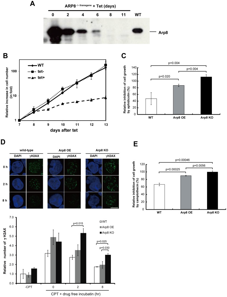
Figure 6. Characterization of Arp8-knockout cells.
(A) Whole-cell extracts were prepared from same number of wild-type (WT) and ARP8 -/-/transgene cells at the indicated times after the addition of 2 µg/ml of tet, and subsequently they were analyzed by Western blot using an anti-Arp8 antibody. (B) Representative growth curves for the WT and ARP8 -/-/transgene cells with (tet+) or without (tet−) tetracycline treatment. Results shown are using cells from day 7 to day 13 after the addition of tetracycline. The number of living cells was counted after trypan blue staining and represented as fold increase in cell number. (C) Sensitivity of WT, Arp8 OE and Arp8-knockout (Arp8 KO; ARP8 -/-/transgene cells cultured in the presence of tet for 8 days) cells to aphidicolin. Cells were cultured in the absence or presence of 0.25 µM aphidicolin for 48 h. The relative inhibition of increase in cell number by aphidicolin (%) was calculated as follows: [{(cell number at 48 h – cell number at 0 h) in the absence of aphidicolin – (cell number at 48 h – cell number at 0 h) in the presence of aphidicolin} ×100]/[(cell number at 48 h – cell number at 0 h) in the absence of aphidicolin]. If the cells did not grow at all in the presence of aphidicolin, then the (cell number at 48 h – cell number at 0 h) in the presence of aphidicolin becomes zero and the relative inhibition becomes 100%. However, sometimes in the presence of aphidicolin there were less number of cells at 48 h than at 0 h (instead of an increase in cell number), in which case the (cell number at 48 h – cell number at 0 h) becomes negative and the relative inhibition becomes more than 100%. (D) Comparison of γ-H2AX foci in wild-type, Arp8 OE, and Arp8 KO cells. The cells were treated with camptothecin (CPT) for 1 h, and after washing out the reagent, the cells were incubated without CPT for 2 h or 8 h. Immunostained γ-H2AX foci were observed under a fluorescence microsope, and the number of foci was counted using the ImageJ software. Plot below shows the number of γ-H2AX foci in the indicated cells relative to that in the camptothecin-untreated (-CPT) wild-type cells. (E) Wild-type, Arp8 OE, and Arp8 KO cells were treated with 1 µM camptothecin for 1 h. After washing out the reagent, the relative inhibition by camptothecin was shown as in C. Error bars indicate average mean ± SD (n = at least 3 independent experiments).

