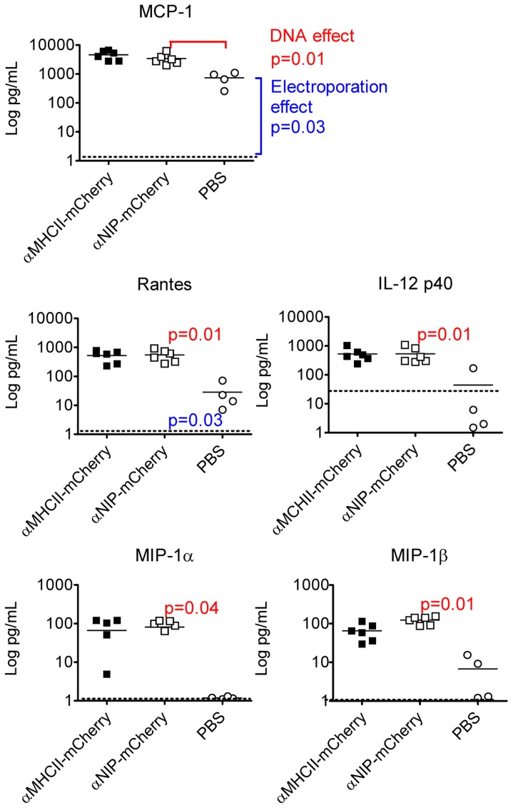
Figure 6. Cytokines in the muscle following intramuscular injection of DNA and EP.
BALB/c were immunized in quadriceps with either PBS or DNA encoding αMHCII-mCherry or αNIP-mCherry, as indicated. After 3 days, muscles from DNA-injected (n = 6/group) and PBS-injected (n = 4/group) were harvested and homogenized before analysis by use of Bio-Plex. Baseline levels, indicated by stippled horizontal lines, represent values measured in muscle isolated 21 days after injection of PBS and electroporation (n = 4). Each symbol represents data from one mouse. Mean values are indicated by horizontal lines. Mann-Whitney test was used to calculate p-values.

