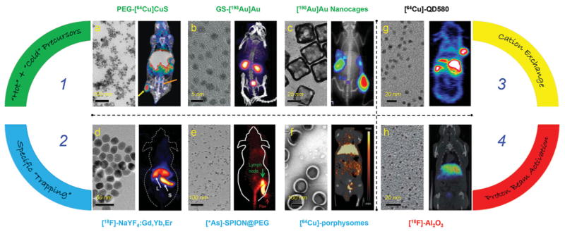
Figure 1.

Representative transmission electron microscopy (TEM) (left panel) and in vivo PET/SPECT (right panel) images of different intrinsically radiolabeled nanoparticles prepared through four different methods. Route 1: Hot-plus-cold precursors. a) TEM image of PEG-[64Cu]CuS nanoparticles and PET/CT image of U87MG xenograft mouse at 24 h p.i. Tumor and bladder were marked with yellow and orange arrows, respectively. Reproduced with permission.[15] Copyright 2010, American Chemical Society. b) TEM image of GS-[198Au]Au nanoparticles and SPECT/CT image of mouse acquired 10 minutes after injection. Reproduced with permission.[19] Copyright 2012, WILEY-VCH Verlag GmbH & Co. KGaA, Weinheim. c) TEM image of [198Au]Au nanocages and 198Au induced Cerenkov luminescence image in EMT-6 tumor bearing mice at 24 h p.i. Reproduced with permission.[20] Copyright 2013, American Chemical Society. Route 2: Specific trapping. d) TEM image of [18F]-NaYF4:Gd3+/Yb3+/Er3+ and whole-body micro-PET image taken 15 minutes p.i. The arrows point at liver (L) and spleen (S). Reproduced with permission.[30] Copyright 2011, Elsevier. e) TEM image of water soluble SPIONs and lymph node PET imaging with *As-SPION@PEG (* = 71, 72, 74, 76) at 2.5 h p.i. Green and red arrows point to lymph node and paw, respectively. Reproduced with permission.[33] Copyright 2013, WILEY-VCH Verlag GmbH & Co. KGaA, Weinheim. f) TEM image of porphysomes and micro PET/CT image in orthotopic PC3 tumor using [64Cu]-porphysomes at 24 h p.i. White arrow points to prostate tumor. Reproduced with permission.[36] Copyright 2012, WILEY-VCH Verlag GmbH & Co. KGaA, Weinheim. Route 3: Cation exchange. g) TEM image of [64Cu]-QD580 and coronal PET image of [64Cu]-QD580 in U87MG bearing mouse at 17 h p.i. Reproduced with permission.[40] Copyright 2014, American Chemical Society. Route 4: Proton beam activation. h) TEM image of [18 F]-Al2O3 nanoparticles and PET/CT fused image at 60–80 min p.i. Reproduced with permission.[44] Copyright 2012, American Chemical Society.
