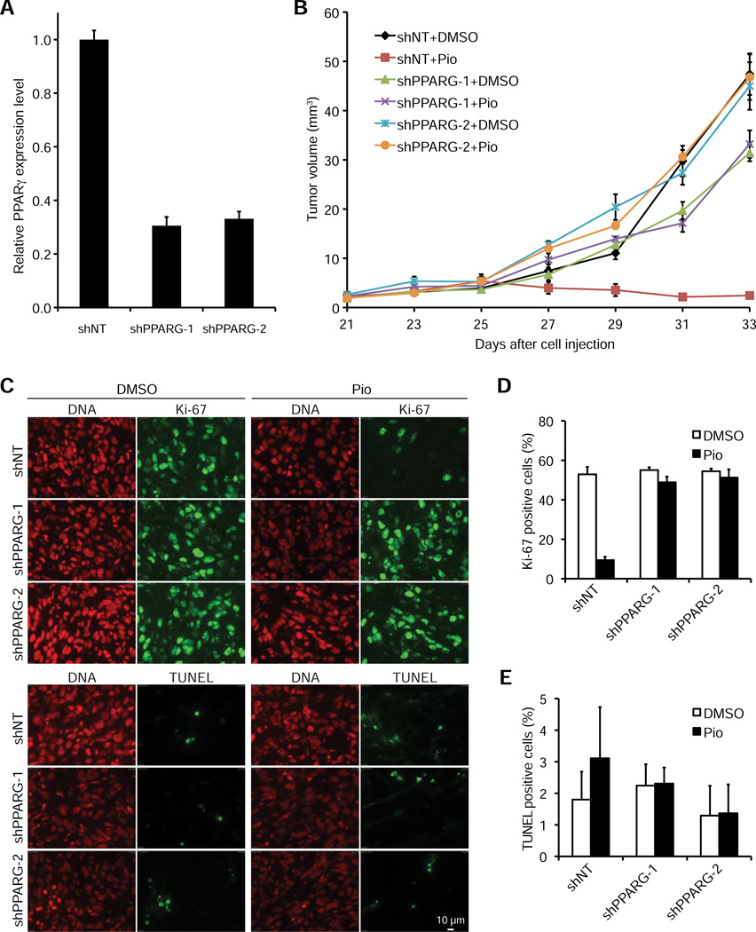
Figure 4. The antitumorigenic effect of pioglitazone in vivo is caused by inhibition of cell proliferation and is PPARγ-dependent.
(A) PPARγ mRNA expression in NCI-H2347 cells stably expressing shNT (negative control), shPPARG-1 or shPPARG-2. PPARγ transcript levels were determined by quantitative RT-PCR using B2M as a calibrator (n=3, error bars represent s.d.).
(B) Mice xenografted with NCI-H2347 shNT, shPPARG-1 and shPPARG-2 cells were treated upon appearance of tumors with DMSO (vehicle) or pioglitazone every other day beginning on day 25 after cell injection. Error bars represent s.e.m. N = 10 for all treatment groups. Growth of the shNT tumors was inhibited by pioglitazone treatment (p<1e-5), but no such effect was observed for the shPPARG-1 and shPPARG-2 tumors (p>0.05).
(C-E) Tumors were obtained at the end of the treatment course and stained for Ki-67, TUNEL, and DNA (propidium iodide) (Scale bar is 10 µm) (C). For quantification of Ki-67 positivity (D) and TUNEL positivity (E) two microscopic fields containing at least 100 cells each were analyzed for each treatment group. Error bars represent s.d.

