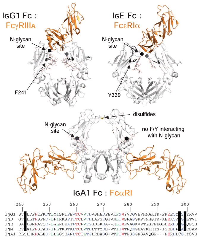
Figure 7. N-glycosylation and an aromatic residue at a comparable position are conserved features of IgD, E, G an M.
Of the human immunoglobulins, only IgA does not share these features, however, the IgA1:FcαRI interaction is remarkably different. The structure of receptor (orange ribbon): Fc (white ribbon) complexes is shown based (pdb: 1e4k (Sondermann et al., 2000) pdb: 1f6a (Garman et al., 2000) pdb: 1ow0(Herr et al., 2003)). At the bottom an amino acid alignment, based on IgG1 Fc numbering, is highlighted to show conserved aromatic residues and N-glycosylation sequons.

