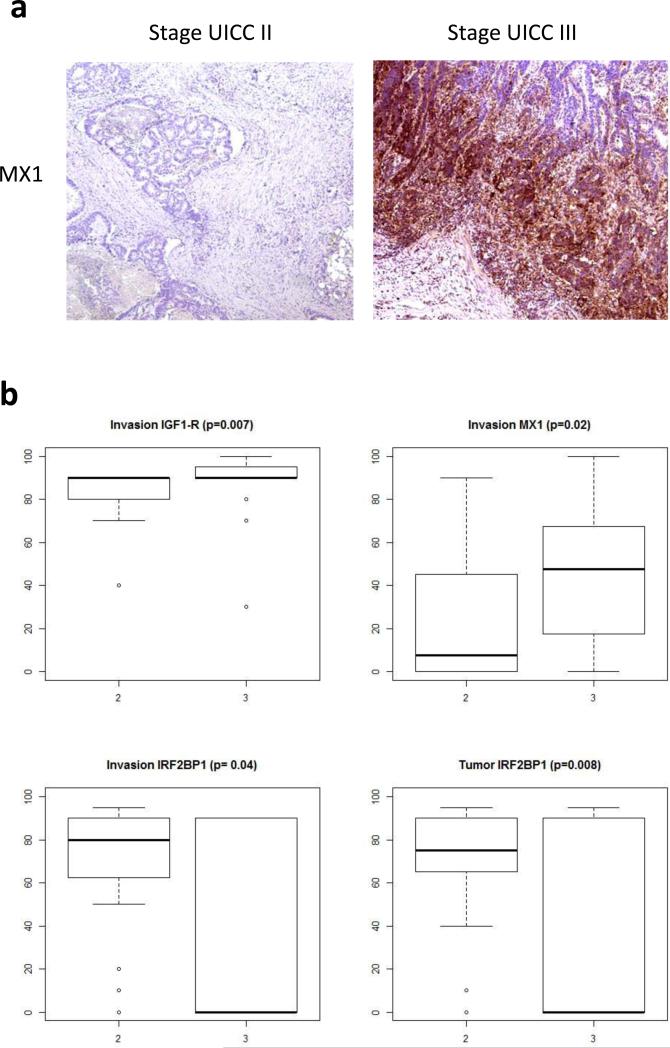
Figure 3.
Validation of candidate proteins by IHC. A: Representative IHC staining of MX1 in stage UICC III and stage UICC II tumors. B: Boxplots for MX1, IGF1-R, and IRF2BP1. The p values calculated by the two-sample Wilcoxon test are indicated for the significant proteins. The validation cohort comprised 20 stage UICC III samples and 20 stage UICC II samples.

