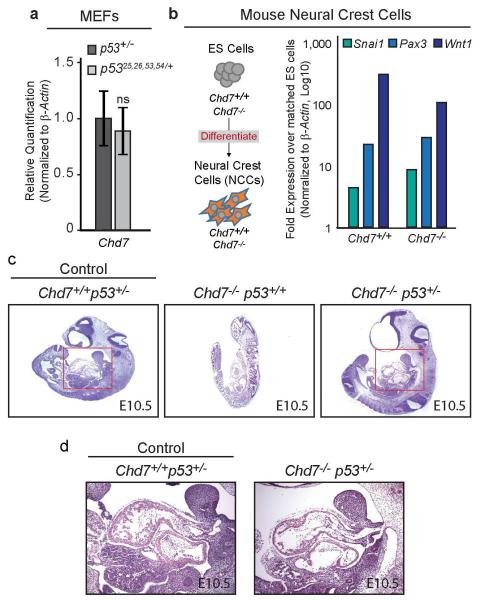
Extended Data Figure 8. p53 Heterozygosity Partially Rescues Chd7-Null Embryos.
(a) qRT-PCR analysis of Chd7 in untreated MEFs derived from E13.5 p53+/− and p5325,26,53,54/+ embryos. Graphs indicate averages from four independent MEF lines, +/−s.d., after normalization to β-actin. ns=non-significant. (b) Left: Schematic of neural crest cell differentiation. Right: Representative qRT-PCR analysis of neural crest cell markers in neural crest-like cells differentiated from Chd7+/+ and Chd7−/− (whi/whi) mouse embryonic stem cells normalized to β-actin and compared to matched embryonic stem cells. (c) H&E-stained E10.5 Chd7+/+p53+/− (control), Chd7−/−p53+/+, and Chd7−/−p53+/− embryos. The Chd7−/−p53+/+ embryo shown is necrotic as evidenced by cellular autolysis. (d) Close-up image of heart region, denoted by red box in panel c, in E10.5 Chd7+/+p53+/− (control) and Chd7−/−p53+/− embryos.

