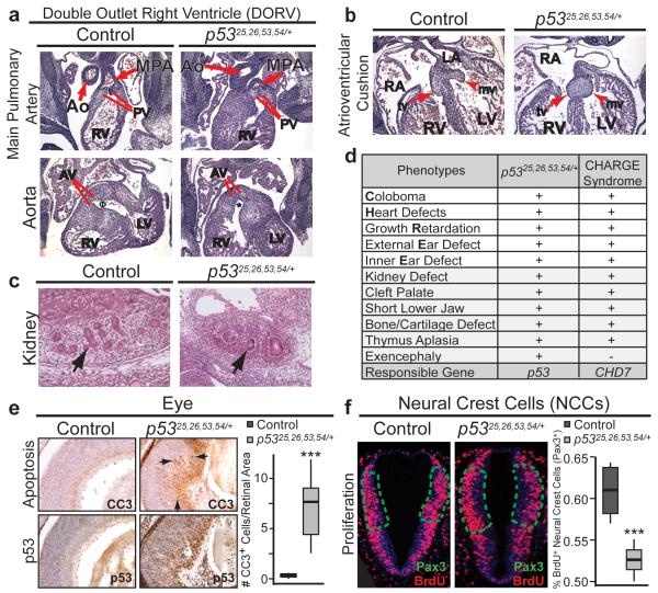
Figure 2. p5325,26,53,54/+ Embryos Exhibit Additional Features of CHARGE Syndrome and p53-Dependent Cellular Responses.
(a) Double outlet right ventricle (DORV) in E13.5 p5325,26,53,54/+ heart (50%, n=6). Top: Main pulmonary artery (MPA) connects via pulmonary valve (PV) to right ventricle (RV) in both control and p5325,26,53,54/+ embryo. Bottom: Aorta (Ao) in control embryo connects to left ventricle (LV) via aortic valve (AV)Φ. Aorta in p5325,26,53,54/+ embryo connects to RV via AV*. (b) Abnormal atrioventricular cushions in E13.5 p5325,26,53,54/+ heart (75%, n=4) fail to elongateinto mature mitral (mv, arrowhead) and tricuspid (tv, arrow) valves. RA: right atrium; LA: left atrium. (c) E13.5 p5325,26,53,54/+ kidneys are smaller (79%), with fewer average glomeruli (13 vs. 3; n=5; arrows), than controls. (d) p5325,26,53,54/+ embryonic phenotypes observed in CHARGE (+present, −absent). (e) Left: Cleaved-caspase 3 (CC3; Top) and p53 (Bottom) immunohistochemistry in E15.5 retinas. Arrows: CC3-positive cells. Right: CC3-positive cells per retinal area. ***p-value=0.007; one-tailed Welsh’s t-test (n=5). (f) BrdU immunofluorescence in E9.5 Pax3+ NCCs (delineated by green-dotted line; Extended-Data Fig. 6c). Right: Percentage BrdU-positive cells per total Pax3+ NCCs ***p-value=0.004 one-tailed Student’s t-test (n=4).

