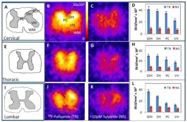
Figure 2. In Vitro Studies on the Rat Spinal Cord with 18F-Fallypride.
20 μm slices of the cervical, thoracic and lumbar rat spinal cord sections from rats injected with 18F-fallypride (n=6). Figures A, E, I are drawings of the cervical, thoracic, lumbar sections respectively, which illustrate the gray matter and white matter of each section. Figures B, F, and J show 18F-fallypride binding in vitro in corresponding sections. Figures C, G, K show nonspecific binding with 10 μM of sulipride in corresponding adjacent sections. Figures D, H, L show total binding and nonspecific binding in the various spinal cord regions (SDH: Superficial dorsal horn; DH: Dorsal horn VH: Ventral horn; PC: Pars centralis; WM: White matter).

